

Table of Contents
Article Summary:
Milia, or "milk spots", are very small white lumps that develop in the layer of skin right underneath the outermost layer. These are benign (not harmful) and occur frequently among infants; they may also develop in children and adults. Most often, milia go away on their own in a couple of weeks without needing medical treatment. There are many ways to get rid of milia professionally if they continue longer than a couple of weeks or if you'd simply like to remove them before they disappear. The following article offers a complete overview regarding what milia are, how they form and grow, and the various methods of treating these small, non-cancerous growths.
Understanding Milia: What Are These Tiny White Bumps?
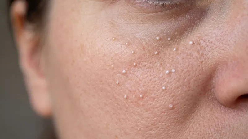
Milia are tiny white or yellowish-coloured cysts located immediately beneath the outermost layer of skin. They are occasionally termed 'oil seeds' or 'milk spots'. ' When there is one cyst, it's called a milium. When there are multiple cysts, it is called milia. While milia primarily develop on the face, they can also be found in other areas of the body.
Milia are not acne. Although they look similar to whiteheads, they are actually quite different. Milias are not transmissible. Therefore, you cannot catch them from someone else. And although they usually don't hurt and are generally free from discomfort for most people, it is possible for them to be painful and uncomfortable in some less typical cases. Approximately 40-50 per cent of newborns enter the world with milia. Therefore, it is one of the most prevalent skin problems in infants. Adult individuals can also develop milias at virtually any point during adulthood.
Identifying the Signs: Common Milia Symptoms
As stated above, milia exhibit a variety of recognisable traits which make identification relatively easy. Some of these features include:
-
Small (typically 1-2 mm) white or yellow bumps on your skin
-
Bumps appearing individually or in large clumps
-
In the majority of cases, the bumps will not be accompanied by any level of pain, redness, itching, etc.
-
Bumps lie directly beneath the skin surface.
Milia are typically seen on your eyelids, underneath your eyes, on your cheeks, forehead, nose, arms, legs, chest, genital area, and even internally in the mouth. The presence of milia will only impact your outward appearance and pose absolutely no risk to your health.
Why Do They Form? Primary Milia Causes
They are caused by dead skin cells becoming trapped under the outer layer of your skin instead of sloughing off normally. As new layers of skin develop above these trapped cells, they harden into cysts. Dead skin cells can be trapped under the skin due to a wide range of factors, including but not limited to:
-
Skin irritation or trauma from burns, blisters, rashes, or extended sun exposure.
-
The prolonged application of steroid creams or ointments on the skin clogs the skin's ability to shed its cells due to heavy applications of topical facial creams and ointments.
-
Autoimmune responses
-
Genetic disorders
While in newborns, the formation of milias is a normal adaptation to life outside the uterus, there does not seem to be any external stimulus required for milias to form. Additionally, since milia are considered a common aspect of early skin development among newborns, there is no specific reason why they should be expected to develop in newborns.
Types of Milia: From Neonatal to Secondary Milia
There are many distinct kinds of milia; however, they are categorised based upon their characteristics and causative agents.
-
Neonatal milia are the most frequent kind of milia. Among all newborns, approximately half will have neonatal milia. Cysts begin forming at birth. Typically, they develop in the nose region. In general, they go away within a few weeks.
-
Primary milias affect both children and adults and generally occur in areas such as the eyelids, forehead, cheeks or genital regions. Like neonatal milia, primary milia will generally go away without treatment.
-
Secondary milia (traumatic milia) develop as a direct result of prior injury to the skin. Damage to the skin from burn injuries, blisters, rashes, and or chronic exposure to sunlight and chemical reactions resulting from heavy use of skin products can all lead to secondary milia.
-
Juvenile milias are associated with inherited genetic diseases. Juvenile milias may first appear at birth or sometime later in childhood.
Milia en plaque is an uncommon kind of milia. It mostly occurs in middle-aged women (between 40 and 60 years old). Clusters of cysts develop on elevated patches of skin behind the ears, along the jawline, near the eyelid, or elsewhere.
Eruptive milias can produce numerous clusters of cysts at varying rates and durations (weeks/months) on the face, upper arms and upper abdominal regions. Eruptive milia can be itchy.
The Clinical Approach: Milia Diagnosis by Professionals
Physicians have an uncomplicated time diagnosing milia and generally do not need to run any diagnostic tests for patients exhibiting classic symptoms. A physician can establish the diagnosis via a routine observation of the affected area. This is especially true for neonatal milia since a biopsy is almost always unnecessary.
When physicians desire to eliminate alternative diagnoses or verify whether their patient's presentation is unusual or persists beyond an expected period of resolution, a biopsy may be taken. Biopsies involve removing a small tissue sample from the affected area for microscopic evaluation. Biopsy is never truly necessary for standard presentations of milia; however, it can be beneficial in identifying unusual or chronic presentations.
Effective Solutions: Professional and Topical Milia Treatment
Milia do not require treatment since they will naturally resolve on their own without any complications. However, if you experience either persistent or worsening lesions or simply prefer to remove them earlier for aesthetic purposes, there exist several viable alternatives for treatment.
Methods used by professionals include:
-
Fine needle extraction by a healthcare provider to surgically remove individual cysts
-
Cryotherapy to freeze and remove individual cysts
-
Tretinoin cream prescriptions to promote the shedding of new skin cells
-
Topical prescription creams or the oral antibiotic minocycline for milia en plaque
Home remedies to assist with healthier-looking Skin include:
-
Daily washing of your face using warm water and mild soap, followed by gently drying your face
-
Application of over-the-counter adapalene gel
-
Regularly protecting your skin from sun damage
-
Using gentle exfoliants that fit your skin type
Never attempt to forcefully express (squeeze) or manually remove a milia lesion yourself. Doing so can lead to potential scarring or infection. Adults desiring faster removal of milia should consult with a healthcare provider or dermatologist rather than attempting removal themselves. Infants should not apply adult moisturisers or oils, as their sensitive infant skin cannot tolerate such applications.
Prevention Tips: How to Minimize Future Milia
Since most cases of milia cannot be avoided, especially neonatal milia, you can lower your chances of developing certain types as an adult by adopting the following preventative behaviours:
-
Reducing prolonged periods of excessive sun exposure and practising regular sun protection techniques.
-
Avoiding prolonged use of thick facial creams and ointments that can block dead skin cells from being shed naturally.
-
Using steroid creams only according to your healthcare provider's instructions.
-
Practising gentle exfoliation or utilising mild cleansing agents to enhance cell turnover.
While adoption of these practices will not ensure prevention, they will significantly minimise your chances of developing additional milia resulting from damage to your skin or clogged pores.
When to Seek Dermatological Advice
In nearly every case, regardless of age, milia do not necessitate a visit with a specialist. However, you may consider consulting with a dermatologist if:
-
Milia has failed to resorb after 2-3 months.
-
Milia continues to worsen, expand, and develop rapidly.
-
Your concern with your appearance prompts you to seek professional removal.
-
A baby's milia has not disappeared after 2-3 weeks post-birth.
Dermatologists can determine the type(s) of milia that you have developed and provide recommendations for removal procedures based on their severity and location.
Conclusion
Milia represent a common benign skin condition experienced by people across the full spectrum of ages, from newborns to elderly adults. In nearly all cases, milia clear up on their own without any medical interventions within 2-3 weeks. Adopting good daily hygiene practices, protecting your skin from excessive UV radiation, and refraining from using excessively rich skincare products can greatly minimise your likelihood of developing specific types of milia. Should your milia remain persistent, progress negatively or if you would like to have them removed sooner for cosmetic reasons, there exist proven treatments available for removal by professionals. Contact a healthcare provider if you have any questions concerning any aspects related to your baby's or your own milia.
References
- Berk, D. R., & Bayliss, S. J. (2008). Milia: A review and classification. Journal of the American Academy of Dermatology,59(6),1050–1063. https://doi.org/10.1016/j.jaad.2008.07.034
- Cutaneous Medicine and Surgery. (2003). Neonatal skin and skin disorders. Dermatologic Clinics, 21(3), 365–374. https://doi.org/10.1016/S0733-8635(03)00031-X
- Horii, K. A., & Drolet, B. A. (2010). Common neonatal dermatological conditions. NeoReviews, 11(9), e499–e509. https://doi.org/10.1542/neo.11-9-e499
- Kolar, S. S. N., & McDaniel, D. H. (2016). Topical retinoids in the management of photodamaged skin. Journal of Drugs in Dermatology, 15(2), 96–102. https://pubmed.ncbi.nlm.nih.gov/26885787
- Zuber, T. J. (2002). Minimal excision technique for epidermoid (sebaceous) cysts. American Family Physician, 65(7), 1409–1412. https://www.aafp.org/pubs/afp/issues/2002/0401/p1409.html

